Anatomy Of Chest X Ray : Normal chest x-ray: Anatomy tutorial | Kenhub / In fact every radiologst should be an expert in chest film reading.. Abcde aproach comparison of pa vs. Gillian lieberman forthe harvard 62. Labeled chest radiographs teaching radiologic anatomy with a level of detail appropriate for medical students. In fact every radiologist and pulmonary physician should be an expert in chest film reading. Submitted 1 year ago by gmdmd.
Evaluation of a chest radiograph may appear to be simple, but is in fact a complex task requiring careful observation, sound understanding of chest anatomy, and knowledge of the principles of physiology and pathology. In this article we will focus on: Common symptoms that can be diagnosed using chest. Therefore, knowing the basics and pathologies in the ed setting is very important. This imaging method can also check how a patient is responding to specific treatments.
Normal Anatomy | Radiology Key from radiologykey.com Presence of metallic objects within the area of examination. Labeled chest radiographs teaching radiologic anatomy with a level of detail appropriate for medical students. Doctors use them to diagnose problems. Both lungs should be well expanded and similar in volume. In fact every radiologst should be an expert in chest film reading. It first appears too complicated to read the chest xrays because we barely know what. It is almost always the first imaging study ordered to evaluate for pathologies of the thorax, although further diagnostic imaging, laboratory tests. Each of these anatomical structures should be viewed using a systematic approach.
Look for lung and pleural pathology.
Presence of metallic objects within the area of examination. The chest exam is performed more frequently than any other exam in the imaging department. Evaluation of a chest radiograph may appear to be simple, but is in fact a complex task requiring careful observation, sound understanding of chest anatomy, and knowledge of the principles of physiology and pathology. In this article we will focus on: The interpretation of a chest film requires the understanding of basic principles. Submitted 1 year ago by gmdmd. It is almost always the first imaging study ordered to evaluate for pathologies of the thorax, although further diagnostic imaging, laboratory tests. A free large database of high quality radiology cases with differential diagnoses and mnemonics to help with board. In fact every radiologst should be an expert in chest film reading. L the portion of the left lung that corresponds anatomically to the right middle lobe is incorporated into the left upper lobe. L these two lobes are separated by a major fissure, identical to that seen on the right side, although often slightly more inferior in location. It first appears too complicated to read the chest xrays because we barely know what. The interpretation of a chest film requires the understanding of basic principles.
Is there any inhaled foreign body? In this article we will focus on: Clinicalchest xray anatomy labeled clinical (i.redd.it). Structure and function of the shoulder complex. Labeled chest radiographs teaching radiologic anatomy with a level of detail appropriate for medical students.
Chest X-Ray Interpretation for Android - APK Download from image.winudf.com Structure and function of the shoulder complex. A free large database of high quality radiology cases with differential diagnoses and mnemonics to help with board. Abcde aproach comparison of pa vs. Major structures are shown in fig. You have completed this module. This imaging method can also check how a patient is responding to specific treatments. In this article we will focus on: L these two lobes are separated by a major fissure, identical to that seen on the right side, although often slightly more inferior in location.
Presence of metallic objects within the area of examination.
It first appears too complicated to read the chest xrays because we barely know what. Doctors use them to diagnose problems. In fact every radiologist and pulmonary physician should be an expert in chest film reading. The interpretation of a chest film requires the understanding of basic principles. Heart and great vessels — assessment of the cardiovascular anatomy includes assessment of heart and chamber size as well as the position and size of the great. Major structures are shown in fig. Submitted 1 year ago by gmdmd. Structure and function of the shoulder complex. In fact every radiologst should be an expert in chest film reading. Many clinical conditions can be evaluated by this simple radiology test. A collection of anatomy notes covering the key anatomy concepts that medical students need to learn. You have completed this module. Common symptoms that can be diagnosed using chest.
Both lungs should be well expanded and similar in volume. Heart abnormalities, including fluid around the heart (pericardial effusion), an enlarged heart (cardiomegaly), heart failure, or abnormal anatomy of the heart can be. This imaging method can also check how a patient is responding to specific treatments. Many clinical conditions can be evaluated by this simple radiology test. Xray is a type of radiography and most widely used investigation.
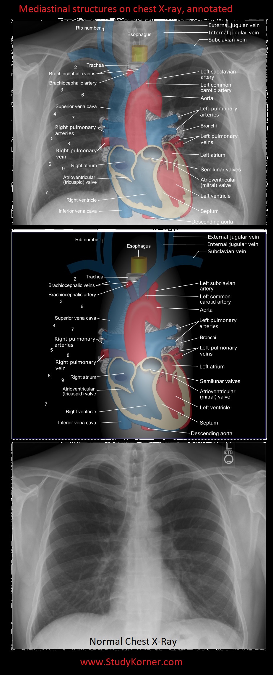
Mediastinal structures on a chest X-ray, annotated - NCLEX ... from www.nclexquiz.com L the portion of the left lung that corresponds anatomically to the right middle lobe is incorporated into the left upper lobe. Common symptoms that can be diagnosed using chest. Labeled chest radiographs teaching radiologic anatomy with a level of detail appropriate for medical students. In fact every radiologist and pulmonary physician should be an expert in chest film reading. A free large database of high quality radiology cases with differential diagnoses and mnemonics to help with board. The interpretation of a chest film requires the understanding of basic principles. Doctors use them to diagnose problems. Both lungs should be well expanded and similar in volume.
Chest radiographs are the most common film taken in medicine.
A free large database of high quality radiology cases with differential diagnoses and mnemonics to help with board. Both lungs should be well expanded and similar in volume. Presence of metallic objects within the area of examination. Each of these anatomical structures should be viewed using a systematic approach. Heart and great vessels — assessment of the cardiovascular anatomy includes assessment of heart and chamber size as well as the position and size of the great. Chest radiographs are the most common film taken in medicine. Structure and function of the shoulder complex. Many clinical conditions can be evaluated by this simple radiology test. This imaging method can also check how a patient is responding to specific treatments. Conclusion of living anatomy of the chest congratulations! L these two lobes are separated by a major fissure, identical to that seen on the right side, although often slightly more inferior in location. Labeled chest radiographs teaching radiologic anatomy with a level of detail appropriate for medical students. The chest exam is performed more frequently than any other exam in the imaging department.
Legit, i can make out the trachea, aorta, outline of the heart, and the diaphragm anatomy of chest. Is there any inhaled foreign body?


0 Komentar